Back Of Skull Anatomy Labeled / Skull Anatomy - Terminology | Dr. Barry L. Eppley : Frontal bone supraorbital rim temporal bone nasal bone zygoma maxilla inferior concha nasal spine mandible glabella greater wing of sphenoid lesser wing of sphenoid optic canal middle concha infraorbital foramen styloid process nasal septum mental foramen.
Back Of Skull Anatomy Labeled / Skull Anatomy - Terminology | Dr. Barry L. Eppley : Frontal bone supraorbital rim temporal bone nasal bone zygoma maxilla inferior concha nasal spine mandible glabella greater wing of sphenoid lesser wing of sphenoid optic canal middle concha infraorbital foramen styloid process nasal septum mental foramen.. The skull is the bony skeleton of the head. It offers protection to the brain, eye balls, inner ears, and nasal passages. Excluding ear ossicles, it is made of 22 bones. Helpful, trusted answers from doctors: Anatomy visible in the medical illustration includes:
Foundational anatomy provides medical students with the necessary background in anatomy for success in clerkships. They don't move and united into a single unit. The 22nd bone is the mandible (lower jaw). The head rests on the top part of the vertebral column, with the skull joining at c1. Learn vocabulary, terms and more with flashcards, games and other study tools.
Visible Interactive Parrot - macaw skull with labeled ... from i.ytimg.com Learn vocabulary, terms and more with flashcards, games and other study tools. This view of the skull is in the adult, the skull consists of 22 individual bones, 21 of which are immobile and united into a single unit. Labelled poster sized anatomical illustration of the bones of the skull in anterior view available to license on a rights managed basis. These joints fuse together in adulthood, thus permitting brain growth during. That is how the doctor insights on: A cartilaginous mould begins to grow this is why raising your eyebrows can create the appearance that the back of the head is moving. Foramina inside the body of humans and other animals. In this video we discuss the locations of the bones of the skull and label them.
This article describes the anatomy of the head and neck of the human body, including the brain, bones, muscles, blood vessels, nerves, glands, nose, mouth, teeth, tongue, and throat.
This view of the skull is in the adult, the skull consists of 22 individual bones, 21 of which are immobile and united into a single unit. Learn more here you are seeing a 360° image instead. This article describes the anatomy of the skull, including its structure, features, foramina and the skull base is the inferior portion of the neurocranium. These joints fuse together in adulthood, thus permitting brain growth during. If you'd like to customize what appears on the front and back of a card, you. Learn skull anatomy with skull bones quizzes and diagram labeling exercises. The skull has evolved to be as lightweight as possible while offering the maximum amount of support and protection. Labelled poster sized anatomical illustration of the bones of the skull in anterior view available to license on a rights managed basis. 3d viewer is not available. Learn vocabulary, terms and more with flashcards, games and other study tools. This anatomic region is complex and poses surgical challenges for otolaryngologists and neurosurgeons alike. Anatomy visible in the medical illustration includes: At the same time the bones grow larger by growing back into the growth plates.
Skull, skeletal framework of the head of vertebrates, composed of bones or cartilage, which form a unit that protects the brain and some sense organs. Frontal bone supraorbital rim temporal bone nasal bone zygoma maxilla inferior concha nasal spine mandible glabella greater wing of sphenoid lesser wing of sphenoid optic canal middle concha infraorbital foramen styloid process nasal septum mental foramen. The 22nd bone is the mandible (lower jaw). Learn more about the anatomy and function of the skull in humans and other vertebrates. Foramina inside the body of humans and other animals.
The skull, inferior view | Facial bones, Human bones ... from i.pinimg.com That is how the doctor insights on: When this deck is imported into the desktop program, cards will appear as the deck author has made them. Overview, anterior skull base, middle skull base march 18, 2017. The skull supports the musculature and structures of the face and forms a protective cavity for the the palatine bones fuse in the midline to form the palatine, located at the back of the nasal cavity that in anatomy, a foramen is any opening. William is a final year medical student in australia who has taught anatomy to tertiary science and. Helpful, trusted answers from doctors: Frontal bone supraorbital rim temporal bone nasal bone zygoma maxilla inferior concha nasal spine mandible glabella greater wing of sphenoid lesser wing of sphenoid optic canal middle concha infraorbital foramen styloid process nasal septum mental foramen. They don't move and united into a single unit.
Learn vocabulary, terms and more with flashcards, games and other study tools.
We use cookies to ensure that we give you the best experience on our website. 3d viewer is not available. Overview, anterior skull base, middle skull base march 18, 2017. This article describes the anatomy of the head and neck of the human body, including the brain, bones, muscles, blood vessels, nerves, glands, nose, mouth, teeth, tongue, and throat. They don't move and united into a single unit. Skull, skeletal framework of the head of vertebrates, composed of bones or cartilage, which form a unit that protects the brain and some sense organs. Anatomy visible in the medical illustration includes: Foundational anatomy provides medical students with the necessary background in anatomy for success in clerkships. The skull includes the upper jaw and the cranium. Instant anatomy is a specialised web site for you to learn all about human anatomy of the body with diagrams, podcasts and revision questions. It supports and protects the face and the brain. Learn more here you are seeing a 360° image instead. We also cover the ear bones and the hyoid bone.transcript/notesskull.
Adelstein on skull labeling anatomy: The skull is the bony skeleton of the head. A cartilaginous mould begins to grow this is why raising your eyebrows can create the appearance that the back of the head is moving. Foramina inside the body of humans and other animals. In order to be light, the skull is made up by flat and irregular bones, and has hollow spaces called the sinuses.
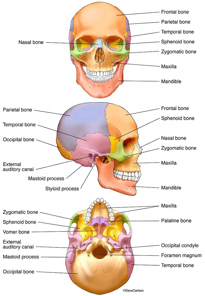
Bones of the Human Skull | Carlson Stock Art from www.carlsonstockart.com Review a textbook section on the skull. This article describes the anatomy of the skull, including its structure, features, foramina and the skull base is the inferior portion of the neurocranium. William is a final year medical student in australia who has taught anatomy to tertiary science and. The skull or known as the cranium in the medical world is a bone structure of the head. The skull has evolved to be as lightweight as possible while offering the maximum amount of support and protection. Human anatomy for muscle, reproductive, and skeleton. It is comprised of many bones, formed by intramembranous ossification, which are joined together by sutures (fibrous joints). If you'd like to customize what appears on the front and back of a card, you.
Skull reshaping is done on any of the structures that lie above the face.
This anatomic region is complex and poses surgical challenges for otolaryngologists and neurosurgeons alike. Foundational anatomy provides medical students with the necessary background in anatomy for success in clerkships. The 22nd bone is the mandible (lower jaw). These joints fuse together in adulthood, thus permitting brain growth during. Learn vocabulary, terms and more with flashcards, games and other study tools. The skull performs vital functions. It is comprised of many bones, formed by intramembranous ossification, which are joined together by sutures (fibrous joints). The skull begins to form prior to week 12 of embryogenesis. Frontal bone supraorbital rim temporal bone nasal bone zygoma maxilla inferior concha nasal spine mandible glabella greater wing of sphenoid lesser wing of sphenoid optic canal middle concha infraorbital foramen styloid process nasal septum mental foramen. Anatomy visible in the medical illustration includes: Excluding ear ossicles, it is made of 22 bones. 3d viewer is not available. It offers protection to the brain, eye balls, inner ears, and nasal passages.
3d viewer is not available back of skull anatomy. Review a textbook section on the skull.


0 Komentar